Sarcomere on Muscle Fiber Model: The Microscopic Engine Behind Every Movement
Sarcomere on Muscle Fiber Model: The Microscopic Engine Behind Every Movement
Beneath the visible surface of human motion lies a microscopic powerhouse: the sarcomere, the functional unit within muscle fibers where mechanical force is born. This precise arrangement of proteins transforms neural signals into coordinated contraction, enabling everything from a sprint to a delicate finger touch. At the heart of this transformation is the sarcomere-on-muscle-fiber model — a framework that elucidates how organized arrays of myosin and actin filaments generate force at the cellular level.
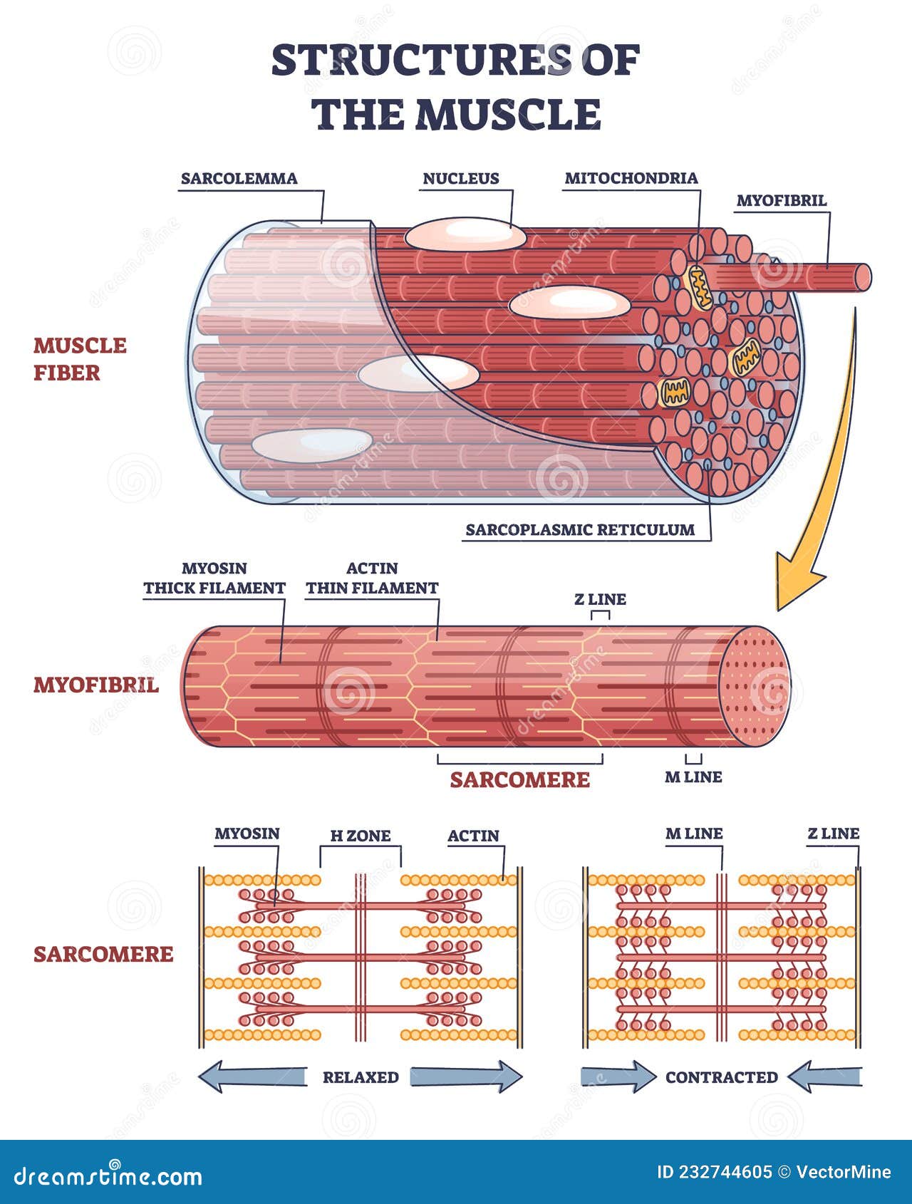
Understanding this model reveals the intricate choreography that makes movement possible, from cellular mechanics to whole-body performance. The sarcomere is not merely a grain of muscle—it is a highly organized structural and functional segment where contraction begins. Composed of interdigitating thin actin and thick myosin filaments arranged in a repeating pattern, the sarcomere serves as the contractile unit of myofibrils within individual muscle fibers.
Each sarcomere contains distinct bands—light and dark—visible under high-resolution microscopy, with Z-lines anchoring the thin filaments at each end. Between them lies the congestion zone rich in myosin, and the M-line stabilizing central components. This precise architecture enables elasticity, strength, and rapid responsiveness.
At the core of muscle contraction lies the sliding filament theory, governed by the sarcomere’s molecular dynamics. When a neuromuscular signal triggers calcium release, myosin heads bind to actin, forming cross-bridges that pivot and pull actin filaments inward, shortening the sarcomere without filament shortening themselves. This process demands ATP and is finely tuned by regulatory proteins like troponin and tropomyosin, which modulate filament access based on calcium levels.
“The sarcomere acts as a molecular machine that converts chemical energy into mechanical work,” explains Dr. Elena Vasilev, molecular biophysicist at the Institute of Muscle Physiology. “Its structural precision ensures force transmission is both efficient and finely regulated.” Visualizing the sarcomere-on-muscle-fiber model reveals hierarchical organization from the ultrastructural to the tissue level.
Within individual muscle fibers, thousands of sarcomeres align end-to-end, forming myofibrils that contribute to the fiber’s overall tensile properties. Bundles of muscle fibers, in turn, integrate into fascicles surrounded by connective tissue, collectively producing the strength and flexibility of actual muscle. Advanced imaging techniques — including cryo-electron microscopy and super-resolution fluorescence microscopy — have illuminated these structures in vivid clarity, reinforcing the model’s predictive power.
This structural blueprint also explains key physiological phenomena: length-tension relationships and force-velocity dynamics. At optimal sarcomere length, maximal actin-myosin overlap allows peak force generation; deviations stretch filaments beyond ideal spacing, reducing efficiency. Similarly, the rate of contraction influences force output—fast—and flexibility governs endurance—slow.
These principles shape muscle performance across diverse activities, from explosive jumps to sustained postures. Beyond basic physiology, the sarcomere model informs clinical and athletic applications. Disorders affecting sarcomere proteins—such as hypertrophic cardiomyopathy and certain myopathies—highlight the consequences of structural breakdown.
Conversely, targeted training and rehabilitation leverage sarcomere adaptability, enhancing hypertrophy, strength, and fatigue resistance through micro-damage and repair cycles. “The sarcomere is the ultimate site of adaptation,” notes Dr. Marcus Reed, sports physiologist at the National Institute of Sports Medicine.
“Understanding its behavior allows us to optimize training, prevent injury, and extend functional lifespan.” Summary| The sarcomere-on-muscle-fiber model illuminates the foundational mechanics of muscle contraction, revealing how nanoscopic protein arrangements generate macroscopic movement. Structured as a precisely aligned network of actin and myosin filaments, the sarcomere operates at the intersection of biochemistry and biomechanics, enabling force generation, elasticity, and dynamic adaptation. From cellular function to whole-body performance, this model remains central to advancing medicine, sports science, and biotechnology.
The sarcomere, visible only through cutting-edge microscopy, is not just a biological unit—it is the very engine of motion, power, and endurance embedded within human anatomy.


Related Post

Ryan Kiera Armstrong: Redefining Soccer’s Next Generation

Revolutionizing Energy: MWSU’s Breakthrough in Molecular Solar Thermal Storage

Al Roker: The Radiant Media Presence Shaped by His Striking Stature

Violet Mom: The Mindful Parenting Movement Redefining Modern Childcare